Frontiers Challenges Associated With the Formation of Protein Inclusion Bodies in
E. coli is a Gram-negative, facultative anaerobic rod that is part of the normal intestinal flora and grows easily in most culture media. E. coli is classified into between 150 and 200 serotypes or serogroups based on somatic (O), capsular (K) and flagellar (H) antigens.
The Natural History of Model Organisms The unexhausted potential of E. coli eLife
E. coli is a facultative (aerobic and anaerobic growth) gram-negative, rod shaped bacteria that can be commonly found in animal feces, lower intestines of mammals, and even on the edge of hot springs. They grow best at 37 C. E. coli is a Gram-negative organism that can not sporulate.

E. coli Symptoms; 14 Warning Signs & Symptoms of E. coli Healthella
Escherichia coli (E. coli) is a gram negative, facultative anaerobic, coliform, rod shaped bacterium It is a part of the normal flora of the intestine of humans and many warm blooded animals; it provides benefits such as production of vitamin K2 and a stable environment where more beneficial bacteria can prosper ( Elife 2015;4:e05826 )

Pin on microbiology
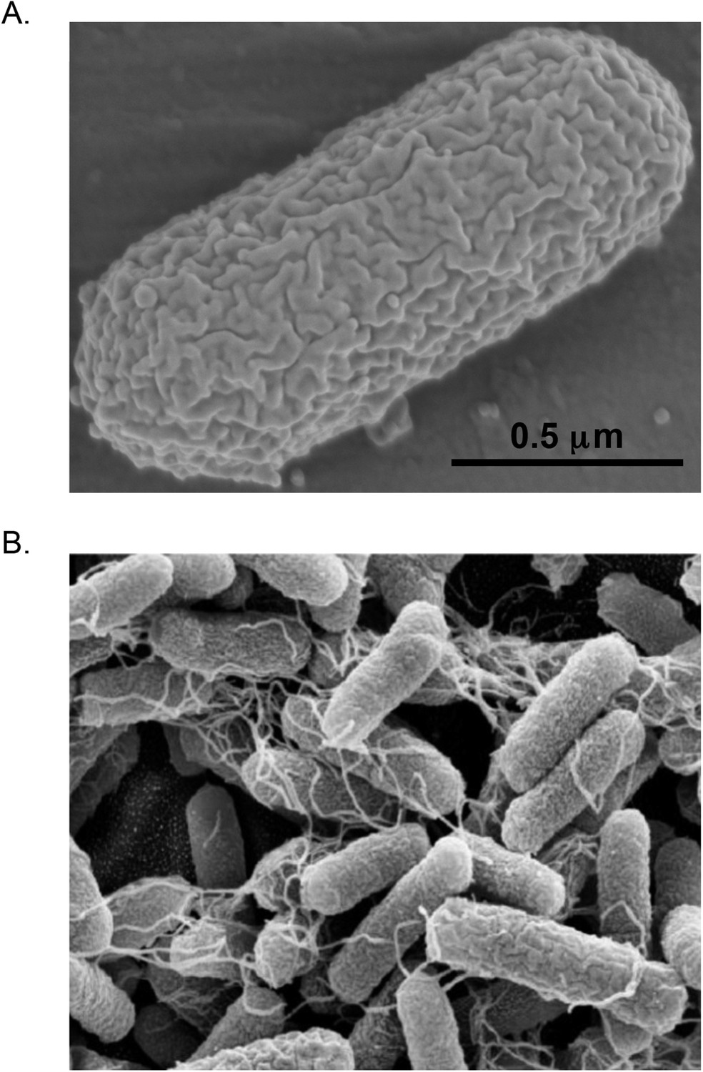
Biology and biochemistry Model of successive binary fission in E. coli Type and morphology E. coli is a gram-negative, facultative anaerobe, nonsporulating coliform bacterium. [18] Cells are typically rod-shaped, and are about 2.0 μm long and 0.25-1.0 μm in diameter, with a cell volume of 0.6-0.7 μm 3. [19] [20] [21]
Prokaryotic Cells
E. coli causes wound infections, usually a result of fecal contamination of external wounds or a result of wounds that cause trauma to the intestinal tract, such as surgical wounds, gunshot wounds, knife wounds, etc. E. coli is by far the most common Gram-negative bacterium causing sepsis. Septicemia is a result of bacteria getting into the.

√100以上 e coli cell diagram 312516E coli cell diagram
E. coli, like most bacteria,. A diagram showing D N A replication. Separated double strand D N A is shown in black. The top black strand runs 3 prime to 5 prime and is attached to a leading strand that is growing from 5 prime to 3 prime due to D N A polymerase moving towards the 3 prime end of the leading strand. The 5 prime end of this.

Cell Cycle Duration Of E Coli Cell Cycle
Researchers uncovered the foundations of biology by using E. coli as a model organism. But over-reliance on this microbe can lead to knowledge blind spots with implications for antibiotic resistance.
Escherichia Coli Stock Illustrations 744 Escherichia Coli Stock Illustrations, Vectors
Escherichia coli is a rod-shaped gram negative bacterium normally resident in human and other mammalian colons. It can grow rapidly on minimal medium that contains a carbon compound such as glucose (which serves both as a carbon source and an energy source) and salts that supply nitrogen, phosphorus, and trace metals.

How the CDC takes on E. Coli, Salmonella, and other foodborne illnesses
To begin drawing an Escherichia coli diagram, you will need a blank sheet of paper or a digital drawing software. Follow these steps to create a basic repres.

Escherichia coli wikidoc
However, optical microscopy studies of single E. coli have been limited by its small size, ∼1 × 3 μ m, not much larger than the optical resolution, ∼0.25 μ m. As a result, not enough quantitative dynamical information on the life cycle of single E. coli is presently available.
bacteria multiplying diagram
Key Facts About Food Poisoning December 1, 2022 , Escherichia coli (abbreviated as E. coli) are bacteria found in the environment, foods, and intestines of people and animals.

Scanning electron microscopy. The morphology of E. coli cells before... Download Scientific
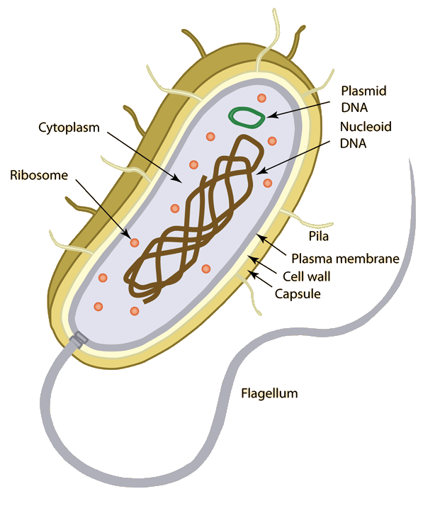
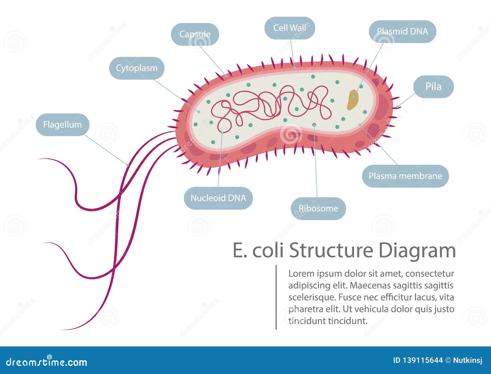
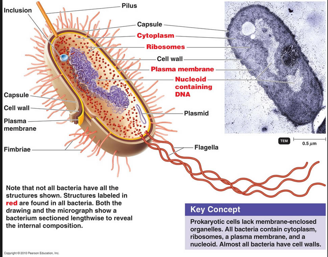
2.2.1 Draw and label a diagram of the ultrastructure of Escherichia coli (E. coli) as an example of a prokaryote. Electron Micrograph of Escherichia coli. 2.2.4 State that bacterial cells divide by binary fission. Binary fission is a form of asexual reproduction and cell division used by prokaryotic organisms.

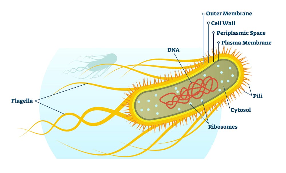
Schematic of E. coli structure and composition, including typical... Download Scientific Diagram
Escherichia coli ( E. coli) is a Gram-negative, rod-shaped, facultative anaerobic bacterium. This microorganism was first described by Theodor Escherich in 1885. Most E. coli strains harmlessly colonize the gastrointestinal tract of humans and animals as a normal flora.
Escherichia Coli Transformation Astral Projection
Escherichia coli is a remarkable and diverse organism. This normally harmless commensal needs only to acquire a combination of mobile genetic elements to become a highly adapted pathogen.
'E. coli' is one of the most widely studied organisms and that may be a problem for both
Home » Bacteriology E. coli (Escherichia coli)- An Overview June 23, 2022 by Sagar Aryal Edited By: Sagar Aryal Table of Contents Habitat of E. coli Morphology of E. coli Antigenic Structure H or Flagellar Antigen O or Somatic Antigen K or Capsular Antigen F or Fimbrial Antigen Cultural Characteristics of E. coli E. coli on Nutrient Agar (NA)

Ultrastructure of Escherichia coli (E. coli) Diagram Quizlet
The haploid circular chromosome in E. coli consists of ~ 4.6 x 10 6 bp. If DNA is relaxed in the B form, it would have a circumference of ~1.5 millimeters (0.332 nm x 4.6 x 10 6) ( Fig 1A ). However, a large DNA molecule such as the E. coli chromosomal DNA does not remain a straight rigid molecule in a suspension.